Most people start searching about pet eye check-ups after noticing something small: a watery eye, a new cloudiness, a bit of redness, a dog squinting in bright light, a cat holding one eye half-closed. Eyes can deteriorate quickly. Some problems are minor irritations, but others are painful emergencies where hours matter.
A good eye exam is not just “a look”. It’s a calm sequence of checks that can spot early disease (before your pet adapts and hides it) and can also rule out urgent problems like corneal ulcers or glaucoma. The sections below lay out what vets look for, what common conditions mean, and what you can sensibly monitor at home.
Why regular eye check-ups matter
Many eye conditions begin quietly. Pets often keep eating, playing and navigating well enough that early vision loss or chronic discomfort is easy to miss. By the time an eye looks obviously “different”, damage may already be advanced.
Routine checks help your vet:
- catch treatable problems early (before scarring or permanent vision loss)
- identify pain (squinting, rubbing, light sensitivity) that owners may interpret as “just tired” behaviour
- monitor age-related change and breed-related risks, especially in dogs with prominent eyes or long facial hair
Common eye problems in pets (and what they usually look like)
Cataracts
Cataracts are an opacity of the lens inside the eye. They often show up as a whitish or grey haze behind the pupil and can reduce vision, sometimes progressing to blindness. A vet exam is important because lens changes can be confused with other age-related cloudiness that looks similar from a distance.
Glaucoma
Glaucoma involves increased pressure inside the eye, which can cause significant pain and rapid, irreversible vision loss. Classic warning signs include a red eye, a cloudy/blue-tinged cornea, tearing, squinting, a dilated pupil, or an eye that looks enlarged. If glaucoma is suspected, it’s treated as an emergency.3
Conjunctivitis
Conjunctivitis is inflammation of the conjunctiva (the tissues lining the eyelids and covering the white of the eye). It often appears as redness, watery to mucky discharge, and irritation (rubbing or pawing). Causes vary: in dogs, ongoing conjunctivitis is commonly linked with allergy and underlying surface disease; in cats, infections such as feline herpesvirus and chlamydia are common contributors.4
What happens in a typical pet eye examination
Most of the core tests are quick and minimally invasive, and they’re chosen to answer practical questions: is the eye producing tears, is the cornea intact, and is the eye pressure safe?
Common steps your vet may use
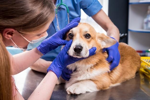
- Visual inspection of eyelids and the surface of the eye for redness, swelling, discharge, lash problems, and obvious injury.
- Schirmer tear test to measure tear production (useful when “dry eye” or chronic irritation is suspected).1
- Fluorescein stain to look for corneal scratches or ulcers and to assess tear drainage. The dye highlights areas where the corneal surface is damaged.1, 2
- Tonometry (eye pressure test) to help detect glaucoma or inflammation such as uveitis.2
Depending on what your vet finds, they may also examine the back of the eye (retina) and lens with an ophthalmoscope, or recommend a referral to a veterinary ophthalmologist for specialised testing and treatment.
When to treat it as urgent
Eye pain and sudden changes should be taken seriously. Book an urgent appointment (or seek an emergency vet) if you notice:
- sudden squinting or holding an eye closed
- cloudiness, a blue/white haze, or a sudden change in eye shape
- a red, swollen, or bulging eye
- a pupil that looks unusually large or unequal
- rapid onset vision changes (bumping into things, hesitation on stairs)
These signs can occur with glaucoma, corneal ulceration, trauma, or uveitis, and delaying care can mean permanent vision loss.3
Fears and misconceptions about eye exams
Most pets tolerate eye checks well. The discomfort, when it happens, is usually from the underlying eye problem rather than the test itself. Your vet will work gently and efficiently, and may use local anaesthetic drops for certain procedures (such as some forms of tonometry).
If your pet is very anxious or painful around the face, tell the clinic at booking. A quieter appointment time, careful handling, or a staged approach can make the visit safer for everyone.
What you can do at home (and what to avoid)
Simple home checks
Once or twice a week—more often for flat-faced breeds or pets with chronic tear staining—take a quick look in good light:
- Are both eyes open comfortably and the same shape?
- Is the surface clear and glossy, not cloudy?
- Is discharge clear and minimal, rather than thick, yellow/green, or bloody?
- Is there new redness, swelling, or frequent rubbing?
Gentle cleaning
If there’s mild discharge at the corner of the eye, wipe it away with a clean, damp cloth or cotton pad, using a separate pad for each eye to avoid spreading infection.
What not to do
- Don’t use human eye drops unless your vet has told you to (some can worsen ulcers or mask serious disease).
- Don’t try to remove a foreign body stuck on the eye surface.
- Don’t “wait it out” when there’s squinting, cloudiness, or obvious pain.
How often should pets have eye checks?
For most pets, an eye assessment is folded into routine veterinary health checks. Pets that are older, have prominent eyes, have had previous eye disease, or belong to predisposed breeds may benefit from more frequent monitoring—your vet can tailor the schedule to what they see on exam.
References
- Merck Veterinary Manual (Professional Version) – Diagnostic tests pertaining to the eye in animals (Schirmer tear test; fluorescein stain)
- Animal Eye Care (Australia) – Eye examination summary (fluorescein staining; tonometry overview)
- Cornell University College of Veterinary Medicine – Glaucoma in dogs (clinical signs; importance of early diagnosis)
- Animal Eye Care (Australia) – Conjunctivitis (common causes and signs in dogs and cats)

Veterinary Advisor, Veterinarian London Area, United Kingdom